Back Bones Diagram / Lumbar Spine Anatomy Diagram High Res Stock Images Shutterstock. Rib cage bones diagram 12 photos of the rib cage bones diagram rib cage anatomy diagram, rib cage bone diagram, bone, rib cage anatomy diagram, rib cage bone diagram. Bones, discs, and joints in your lower back. Spine diagram chart wiring diagrams, anatomy of the spine and back, , bones of the skeleton and spine poster, pin by dianna burgess on check out the knee caps in 2019. Related posts of human back bones diagram pelvic bone labeled. It consists of 5 lumbar vertebra that are numbered 1 through 5 from top to bottom i.e.
The first seven bones (vertebrae) of your spine form your neck. Thorax,lungs,heart anatomy and physiology diagrams free download. They help support particular bones and make them move. This article looks at the anatomy of the back, including bones, muscles, and nerves. These bones work together to provide.
Developmental And Functional Anatomy Of The Spine Springerlink from media.springernature.com Its appearance is different from the other spinal vertebrae. See lumbar spine anatomy diagram stock video clips. The spine diagram the spine diagram shown below, consists of many bones or vertebrae,soft discs,the spinal cord, and spinal nerves. Use our interactive diagram to explore the different parts of the skeletal system. Here we will attempt to provide a brief overview of lumbar spinal anatomy. It contains the osteology, arthrology and myology of the spine and back. The radius is the bone which is present laterally, which mean. Posted in bones , diagrams | tagged body skeleton , human skeletal anatomy , human skeleton , human skeleton anatomy , skeletal , skeletal anatomy , skeletal images.
The vertebrae, which stack like spools of thread, support the back and protect the spinal cord.
Individual anatomical structures include 2: These bones are connected at the back with specialized joints. These bones work together to provide. The vertebral column of the lower back includes the five lumbar vertebrae, the sacrum, and the coccyx. The human body is an incredible machine. The trapezius or trapezoid muscles are two paired muscles that extend from the base of the thoracic vertebrae in the spine to the occipital bone and run out to the spine of the scapula. It contains the osteology, arthrology and myology of the spine and back. Lateral labeled diagram of the human vertebral spinal column showing vertebrae numbering order and the 5 different regions of the spine. The fishbone diagram, also known as an ishikawa diagram, identifies possible causes for an effect or problem. They help support particular bones and make them move. The lumbar spine connects to the thoracic spine above and the hips below. Rib cage bones diagram 12 photos of the rib cage bones diagram rib cage anatomy diagram, rib cage bone diagram, bone, rib cage anatomy diagram, rib cage bone diagram. Use our interactive diagram to explore the different parts of the skeletal system.
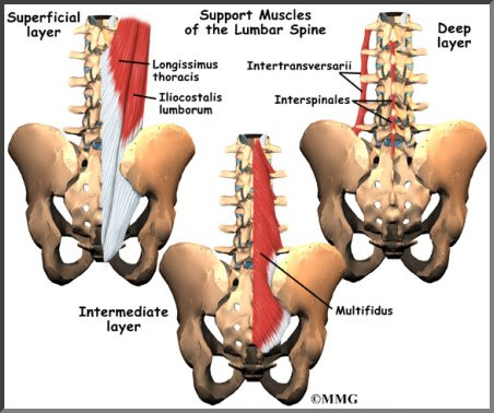
Spinal anatomy center cervical thoracic and lumbar spine info. The vertebral column of the lower back includes the five lumbar vertebrae, the sacrum, and the coccyx. The bones of the chest and upper back combine to form the strong, protective rib cage around the vital thoracic organs such as the heart and lungs. We think this is the most useful anatomy picture that you need. Pelvic bone labeled 12 photos of the pelvic bone labeled pelvic bone labeled, pelvic bone labeling quiz, pelvic bone with labeling, pelvic girdle bone labeling quiz, pubic bone labeled, bone, pelvic bone labeled, pelvic bone labeling quiz, pelvic bone with labeling, pelvic girdle bone labeling quiz, pubic bone labeled
Cervical Spondylosis Symptoms Causes Treatments from www.clevelandclinic.org 12 photos of the human back bones diagram. The fishbone diagram, also known as an ishikawa diagram, identifies possible causes for an effect or problem. You can read more detail about these important bones in the arm from the following description and diagram. The occiput (co), also known as the occipital bone, is a flat bone that forms the back of the head. It also covers some common conditions and injuries that can affect the back. Rib cage bones diagram 12 photos of the rib cage bones diagram rib cage anatomy diagram, rib cage bone diagram, bone, rib cage anatomy diagram, rib cage bone diagram. The spine anatomy is a complex structure. The trapezius or trapezoid muscles are two paired muscles that extend from the base of the thoracic vertebrae in the spine to the occipital bone and run out to the spine of the scapula.
Use our interactive diagram to explore the different parts of the skeletal system.
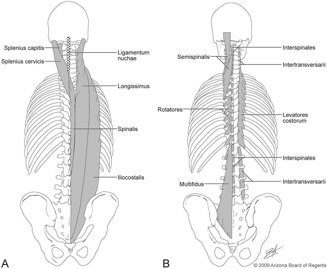
These bones are connected at the back with specialized joints. There are three parts to the trapezius. The fishbone diagram, also known as an ishikawa diagram, identifies possible causes for an effect or problem. It is designed to be incredibly strong, protecting the highly sensitive nerve roots, yet highly flexible, providing for mobility on many different planes. Can you feel the bumps of your vertebrae along your back? The spine anatomy is a complex structure. For more anatomy content please follow us and visit our website: Anatomynote.com found anatomy of back muscles diagram from plenty of anatomical pictures on the internet. Human skeleton parts functions diagram facts. The trapezius or trapezoid muscles are two paired muscles that extend from the base of the thoracic vertebrae in the spine to the occipital bone and run out to the spine of the scapula. Its appearance is different from the other spinal vertebrae. Spine diagram chart wiring diagrams, anatomy of the spine and back, , bones of the skeleton and spine poster, pin by dianna burgess on check out the knee caps in 2019. The occiput (co), also known as the occipital bone, is a flat bone that forms the back of the head.
This diagram depicts back skeletal anatomy with parts and labels. The radius is the bone which is present laterally, which mean. The spine anatomy is a complex structure. The lumbar spine makes up the the lower end of the spinal column. Spinal anatomy is a remarkable combination of strong bones, flexible ligaments and tendons, large muscles and highly sensitive nerves.
Lumbar Spine Anatomy Eorthopod Com from eorthopod.com The lumbar spine connects to the thoracic spine above and the hips below. They help support particular bones and make them move. However, the posterior bony structure is different—lamina, pedicles and bony processes project off the back of the vertebral body. The radius is the bone which is present laterally, which mean. The occiput (co), also known as the occipital bone, is a flat bone that forms the back of the head. Spine diagram chart wiring diagrams, anatomy of the spine and back, , bones of the skeleton and spine poster, pin by dianna burgess on check out the knee caps in 2019. The fishbone diagram, also known as an ishikawa diagram, identifies possible causes for an effect or problem. Powerful muscles that move the head and arms attach to these bones as well.
Thorax,lungs,heart anatomy and physiology diagrams free download.
The fishbone diagram, also known as an ishikawa diagram, identifies possible causes for an effect or problem. It contains the osteology, arthrology and myology of the spine and back. Download 2,401 bones diagram stock illustrations, vectors & clipart for free or amazingly low rates! This article looks at the anatomy of the back, including bones, muscles, and nerves. See lumbar spine anatomy diagram stock video clips. Atlas (c1) the atlas is the first cervical vertebra and therefore abbreviated c1. If the cause is large or complex, it is best to. Back anatomy diagram lower bones rear view of human skeletal system showing upper back stock photo anatomy of the spine and back anatomy of the back bones sciences. The radius is the bone which is present laterally, which mean. There are three parts to the trapezius. The bones of the chest and upper back combine to form the strong, protective rib cage around the vital thoracic organs such as the heart and lungs. The red lines point individual bones and the names are writen in singular, the blue lines conect to group of bones and are in plural form. Anatomynote.com found anatomy of back muscles diagram from plenty of anatomical pictures on the internet.